Saturday, September 22, 2012

Lab 3: Chytridiomycota and Blastocladiomycota motility and morphology


Objective:

To observe sporangiophore and zoospores of Chytridiomycota and Blastocladiomycota.

Method:

Regular mounting method; microscopy.

Result:

1. Sporangiophore of Phytophthora infestans with cell wall staining dye.

Phytophthora is a genus of plant-damaging Oomycetes, whose member species are capable of causing enormous economic losses on crops worldwide, as well as environmental damage in natural ecosystems.

The specialized branches are sporangiophore. Sporangia are attached to sporangiophores. Sporangia are lemon-shaped cells in the picture.
 

2. Sporangia of Allomyces.

Sporangia are enclosed cells which produce zoospores.

3. Relased zoospores from sporangiophore of Allomyces.
Sporagia go through cytoplasmic cleavage to produce many swimming  zoospores. Zoospores are moving fast in a random direction. A zoospore cell is changing shape while it moves. Flagellum can be seen with a certain light reflection angle. Zoospores used flagellum to swim towards a host plant.

Lab 2: Methods of conidiaphore for microscopy


Objective:

To learn different methods for observing aspergelli conidiaphore under microscope.

Methods:

1. Agar block: directly cut a big block of agar from a fungal culture plates and observe conidiaphore under microscope.

2. Riddell mount: put a piece of filter paper on a clean petri dish; wet the filter paper by pouring a little amount of autoclave water in it; put a bend glass rock in petri dish; place a slide on the glass rock; cut a agar block (a little smaller than a cover slide); gentelly place on the silde above the glass rock; inoculate the agar block with a fungal culture; gentelly place a cover slide on the top of agar block; close the petri dish; place in a moisture chamber; incubate at 28 degree for a few days. After the fungal hyphae grow up; gentelly lift the cover slide off the agar block; put on another slide with the hyphae side down; use some glass chips as spacer to avoid squash the conidiaphore.

Result:
1. Agar block method is good for observing fungal hyphae and conidiaphore with intact form. but the defect of this method is it's hard to focus on a single conidiaphore since the sample is thick which only allows to use 10x magnification for observing.


Aspergellus tamari: conidiaphore


Aspergellus sojae: conidiaphore


Aspergellus paraititis: conidiaphore


Aspergellus niger: conidiaphore and aerial hyphae


  Aspergellus nidulans: conidiaphore

 
Aspergellus flavus: conidiaphore
 

2. Riddell mount:

Aspergellus sojae: conidiaphore


Conclusion: both agar block and riddell mount method  are good for fungal conidiaphore observation. However, agar block method can’t get a close good at the conidiaphore since the thickness of the agar block. Riddell mount has a better chance to get a good image of conidiaphore because conidiaphore grow on the surface of the cover slide.

Lab 1 visulization of different fungul species morphology; cell counting with hemocytometer,


Objective:

To Learn different micosopy techniques.

Method:

1.       Squash mount: put a drop of water on the middle of slide; pick a small agar block from fungal culture plate; place on the center of the water drop; cover the agar block with a cover slide; slight twist the cover slide to aside to squash the agar block; observe under microsopce.

2.       Tap mount: put a drop of water on the middle of slide; use a double side tape to touch the surface of a fungal culture; place the tape on the water drop with the specimen side down; directly observe under microsope.

3.       Hemocytometer usage: The coverslip is placed over the counting surface prior to putting on the cell suspension. The suspension is introduced into one of the V-shaped wells with a pasteur or other type of pipet. The area under the coverslip fills by capillary action. Enough liquid should be introduced so that the mirrored surface is just covered. The charged counting chamber is then placed on the microscope stage and the counting grid is brought into focus at low power.

Result and conclusion:
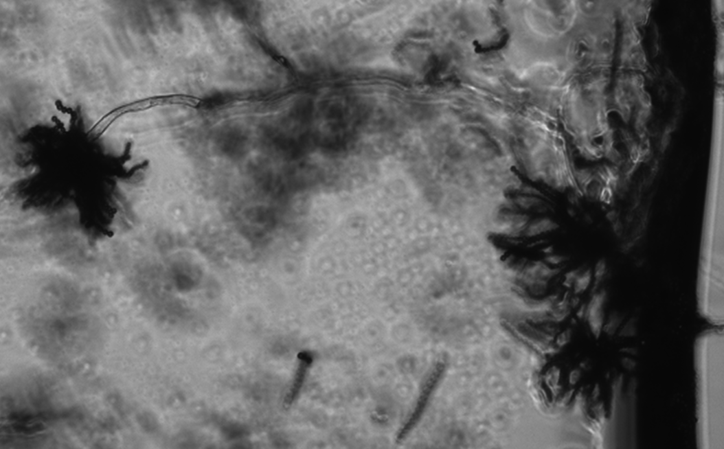
1.       Aspergillus niger: Aspergillus niger is a fungus and one of the most common species of the genus Aspergillus. It causes a disease called black mold on certain fruits and vegetables such as grapes, onions, and peanuts, and is a common contaminant of food. Each cell is a conidia. The usually form a chain.
 
 
2.       Cladosporium: Cladosporium is a genus of fungi including some of the most common indoor and outdoor molds. Species produce olive-green to brown or black colonies, and have dark-pigmented conidia that are formed in simple or branching chains.


3.       Pythium: Pythium is a genus of parasitic oomycete. They are commonly called water moulds. Most species are plant parasites.


4.      Thkelaviopsis bassicola: this fungus forms two types of  spores, the darker one is called aleuriospore; the light color one is called phialospore.
 5. Counting cell number with hemacytometer:  full grid on a hemacytometer contains nine squares, each of which is 1 mm square The central counting area of the hemacytometercontains 25 large squares and each large square has 16 smaller squares. When counting, count only those cells on the lines of two sides of the large square to avoid counting cells twice.
 

Tuesday, September 4, 2012

Self introduction

My name is Xin Chen from China. I am third year graudate student from Plant Path Department. My PI is Dr. Libo Shan. Our lab is interested in studying plant signaling transduction upon pathogen infection.