Objective:
To learn different methods for observing aspergelli conidiaphore under microscope.
Methods:
1. Agar block: directly cut a big block of agar from a fungal culture plates and observe conidiaphore under microscope.
2. Riddell mount: put a piece of filter paper on a clean petri dish; wet the filter paper by pouring a little amount of autoclave water in it; put a bend glass rock in petri dish; place a slide on the glass rock; cut a agar block (a little smaller than a cover slide); gentelly place on the silde above the glass rock; inoculate the agar block with a fungal culture; gentelly place a cover slide on the top of agar block; close the petri dish; place in a moisture chamber; incubate at 28 degree for a few days. After the fungal hyphae grow up; gentelly lift the cover slide off the agar block; put on another slide with the hyphae side down; use some glass chips as spacer to avoid squash the conidiaphore.
Result:
1. Agar block method is good for observing fungal hyphae and conidiaphore with intact form. but the defect of this method is it's hard to focus on a single conidiaphore since the sample is thick which only allows to use 10x magnification for observing.
Aspergellus tamari: conidiaphore

Aspergellus sojae: conidiaphore

Aspergellus paraititis: conidiaphore

Aspergellus niger: conidiaphore and aerial hyphae

Aspergellus nidulans: conidiaphore

Aspergellus flavus: conidiaphore

2. Riddell mount:
Aspergellus sojae: conidiaphore
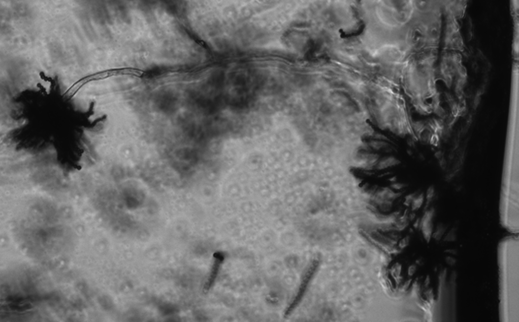
Conclusion: both agar block and riddell mount method are good for fungal conidiaphore observation. However, agar block method can’t get a close good at the conidiaphore since the thickness of the agar block. Riddell mount has a better chance to get a good image of conidiaphore because conidiaphore grow on the surface of the cover slide.
No comments:
Post a Comment