Wednesday, October 24, 2012

Lab 7 Fluoresence microscopy and protein localization; continuation of Neurospora genetics

What's learned
1.Fungal genetic stock center website (www. fgsc. net). The stock center contains all experimental fungal strains. Knockout lines of certain genes can be ordered from the stock center. For example,it provides all information for Neurospora.

2. Knock out a fungal gene: homologous recombination.
This method can directly replace the coding region to block the gene function completely. It has advantage over point mutation.

Objective:
1.Pick single ascospore from mating plate for heat shock.

2.Observe nuclei, actin, tubulin, Mak-2 under fluorescence microspores.

3.Observe mushroom/Chlorophylum sp. basidiospores, Buller's Drop and hyphae clamp connection.
The purpose for this exercise is to practice on picking single ascospores from agar block, and taking video for Neurospora hyphae growth. Additionally,to get a general idea of some subcellular structure localization.

Material and Method:
Material:4% Water agar plate;picker,scaple,slides
Method:
1.Picking random ascospores

  • Use scaple to cut water agar plate into 4cm block and put it on slide
  • Spread loopful of spores-not too many because we want it well seperate
  • Flame picker briefly-the advantage of the picker is it can cool down immediately.
  • Use the picker as scaple to cut out a block with spores
  • Pick up block of agar by scoping`0.2-0.3 mm square
  • Transfer to agar tubes
  • Transfer rack of tubes to refrigerator to allow spores to hydrate.
  • Heat shock:Next day incubate the rack of tubes at 65 degree water bath for 45min.
2. Observer fluorescence signal from Nuerospora mutant.

  • Cut a block of agar which is a little smaller than slide with hyphae tip in the middle. Gently lay the agar block on the slide and lay a long cover slide on the top without push. 
  • Observe fluorescence signal under microscope. 


Result:
1. 10 ascospores are picked from the water agar block and transfer to slide medium agar tubes. Tubes are incubated at room temperature.

2. Observe microtubel tagged with GFP in SMRP11 mutant and nuclei tagged with GFP in SMRP10 mutant.
Under microscope I observe hyphae grow every second. The spizenkoper is wandering
because the hyphae is looking for nutrient source. Time lap is setup to capture each second of hyphae growth.

3. Mushroom/Chlorophylum sp. morphology observation:
Cut gill and observe basidispores.


4. Cut carp to observe hyphae and clamp connection.
Discussion:
From this experiment, I learned how to recognize ascospores and how to use a picker to pick every single ascospore. I also observe basidiospores and clamp connection from mushrooms. The clamp connection observation give me a vivid image of what clamp connection look like in natural condition which help me to understand the lecture better. I think we should do this kind of experiment more. After a lecture of some typical fungal morphogenesis, we are provided a chance to observe what it actually look like other than just get impression from textbook. It will help us to understand the lecture better and leave a longer time memory on the knowledge. 


Tuesday, October 9, 2012

Lab 5 Mitosporic fungi survey

Objective:
Observe conidia morphology of different fungi.
Method:
Regular mounting technique
Results:
1. Botrytis cinera: conidia are borne in grape-like clusters. Conidia are hyaline, ellipsoid to obovoid. There is contamination in Botrytis cinera. There are two conidia forms  present in the view. Based on my study, the small round ones are Botrytis cinera conidia.

2. Curvularia sp.: The poroconidia are curved slightly to distinctly, transversely septate, with an expanded third cell from the pore end of the conidium.

 
3. Epicoccum: Conidia look round, non-septate, brown to black in color, with both transverse and oblique septa, which makes them resemble a soccer ball.

4. Monilinia Fructicola: Conidia are hyaline (colorless), lemon-shaped, and produced in a moniloid manner (resembling a string of beads with constricted ends).
5. Rhizoctonia Solani: it does not produce spores and is hence identified only from mycelial characteristics or DNA analysis. Its hyphal cells are multinucleated. The hyphae are 4–15 μm wide and tend to branch at right angles. A septum near each hyphal branch and a slight constriction at the branch.

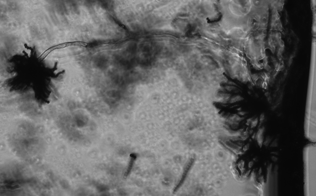
6. Thielaviopsis Basicola:  two types of conidia:
Type 1 conidial chains: terminal, with dark grey walls, smooth, in chains of (2)3-9 so appearing multiseptate, shortly cylindric with flat ends.
Type 2 phialoconidia: hyaline, smooth, cylindric, flat ended, very variable in size with 3-7 septate towards base.


Monday, October 8, 2012

lab 4 Dimorphism and mating: Zygomycete and Basidiomycete

Objective:
Observe the dimorphism for Mucor rouxii; observe ascospore, macroconidia and microconida for Neurospora; make cross for different mating type of Neurospora; inoculate Ustilago to maize seedlings and observe symptom development.
Method:
1. Different conidia form observation: use regular mounting method to observe dimorphism for Mucor rouxii; ascospore, macroconidia and microconida for Neurospora.
2. Cross: wet steriled loop, touch the side of tubes where Neurospora perithecia shot ascospores; inoculate the different mating type strain on opposite side of medium; incubate at room temperature for mating.
3. Ustilago infection:inject diluted  Ustilago spore suspension to maize stem.
Result:
1. Mucor rouxii has dimorphism, it can grow as yeast cell and hyphae. Dimorphism can be observed with a cross section Mucor culture medium. Hyphae will be observed on the upper layer. Yeast cell can be observed on the bottom layer. There are some transition cells in the middle layer.
Hyphae:
Budding yeast cell.

2. Conidiaphore of Neurospora mutant: microconidia don't break from each other.

Ascospore of Neurospora

Macroconidia and microconidia of Neurospora
Macroconidia develop under normal condition, microconidia develop under poor nutrition condition, mostly involved in mating. 

Neurospora mating: different mating type strain hyphae grow toward each other, fuse to produce perithecia along the lines hyphae meet. 


3. Ustilago inoculation: one week after inoculation, there are galls appeared on the leaves.
Galls will develop into lesions with times. 


Saturday, September 22, 2012

Lab 3: Chytridiomycota and Blastocladiomycota motility and morphology


Objective:

To observe sporangiophore and zoospores of Chytridiomycota and Blastocladiomycota.

Method:

Regular mounting method; microscopy.

Result:

1. Sporangiophore of Phytophthora infestans with cell wall staining dye.

Phytophthora is a genus of plant-damaging Oomycetes, whose member species are capable of causing enormous economic losses on crops worldwide, as well as environmental damage in natural ecosystems.

The specialized branches are sporangiophore. Sporangia are attached to sporangiophores. Sporangia are lemon-shaped cells in the picture.
 

2. Sporangia of Allomyces.

Sporangia are enclosed cells which produce zoospores.

3. Relased zoospores from sporangiophore of Allomyces.
Sporagia go through cytoplasmic cleavage to produce many swimming  zoospores. Zoospores are moving fast in a random direction. A zoospore cell is changing shape while it moves. Flagellum can be seen with a certain light reflection angle. Zoospores used flagellum to swim towards a host plant.

Lab 2: Methods of conidiaphore for microscopy


Objective:

To learn different methods for observing aspergelli conidiaphore under microscope.

Methods:

1. Agar block: directly cut a big block of agar from a fungal culture plates and observe conidiaphore under microscope.

2. Riddell mount: put a piece of filter paper on a clean petri dish; wet the filter paper by pouring a little amount of autoclave water in it; put a bend glass rock in petri dish; place a slide on the glass rock; cut a agar block (a little smaller than a cover slide); gentelly place on the silde above the glass rock; inoculate the agar block with a fungal culture; gentelly place a cover slide on the top of agar block; close the petri dish; place in a moisture chamber; incubate at 28 degree for a few days. After the fungal hyphae grow up; gentelly lift the cover slide off the agar block; put on another slide with the hyphae side down; use some glass chips as spacer to avoid squash the conidiaphore.

Result:
1. Agar block method is good for observing fungal hyphae and conidiaphore with intact form. but the defect of this method is it's hard to focus on a single conidiaphore since the sample is thick which only allows to use 10x magnification for observing.


Aspergellus tamari: conidiaphore


Aspergellus sojae: conidiaphore


Aspergellus paraititis: conidiaphore


Aspergellus niger: conidiaphore and aerial hyphae


  Aspergellus nidulans: conidiaphore

 
Aspergellus flavus: conidiaphore
 

2. Riddell mount:

Aspergellus sojae: conidiaphore


Conclusion: both agar block and riddell mount method  are good for fungal conidiaphore observation. However, agar block method can’t get a close good at the conidiaphore since the thickness of the agar block. Riddell mount has a better chance to get a good image of conidiaphore because conidiaphore grow on the surface of the cover slide.

Lab 1 visulization of different fungul species morphology; cell counting with hemocytometer,


Objective:

To Learn different micosopy techniques.

Method:

1.       Squash mount: put a drop of water on the middle of slide; pick a small agar block from fungal culture plate; place on the center of the water drop; cover the agar block with a cover slide; slight twist the cover slide to aside to squash the agar block; observe under microsopce.

2.       Tap mount: put a drop of water on the middle of slide; use a double side tape to touch the surface of a fungal culture; place the tape on the water drop with the specimen side down; directly observe under microsope.

3.       Hemocytometer usage: The coverslip is placed over the counting surface prior to putting on the cell suspension. The suspension is introduced into one of the V-shaped wells with a pasteur or other type of pipet. The area under the coverslip fills by capillary action. Enough liquid should be introduced so that the mirrored surface is just covered. The charged counting chamber is then placed on the microscope stage and the counting grid is brought into focus at low power.

Result and conclusion:

1.       Aspergillus niger: Aspergillus niger is a fungus and one of the most common species of the genus Aspergillus. It causes a disease called black mold on certain fruits and vegetables such as grapes, onions, and peanuts, and is a common contaminant of food. Each cell is a conidia. The usually form a chain.
 
 
2.       Cladosporium: Cladosporium is a genus of fungi including some of the most common indoor and outdoor molds. Species produce olive-green to brown or black colonies, and have dark-pigmented conidia that are formed in simple or branching chains.


3.       Pythium: Pythium is a genus of parasitic oomycete. They are commonly called water moulds. Most species are plant parasites.


4.      Thkelaviopsis bassicola: this fungus forms two types of  spores, the darker one is called aleuriospore; the light color one is called phialospore.
 5. Counting cell number with hemacytometer:  full grid on a hemacytometer contains nine squares, each of which is 1 mm square The central counting area of the hemacytometercontains 25 large squares and each large square has 16 smaller squares. When counting, count only those cells on the lines of two sides of the large square to avoid counting cells twice.
 

Tuesday, September 4, 2012

Self introduction

My name is Xin Chen from China. I am third year graudate student from Plant Path Department. My PI is Dr. Libo Shan. Our lab is interested in studying plant signaling transduction upon pathogen infection.